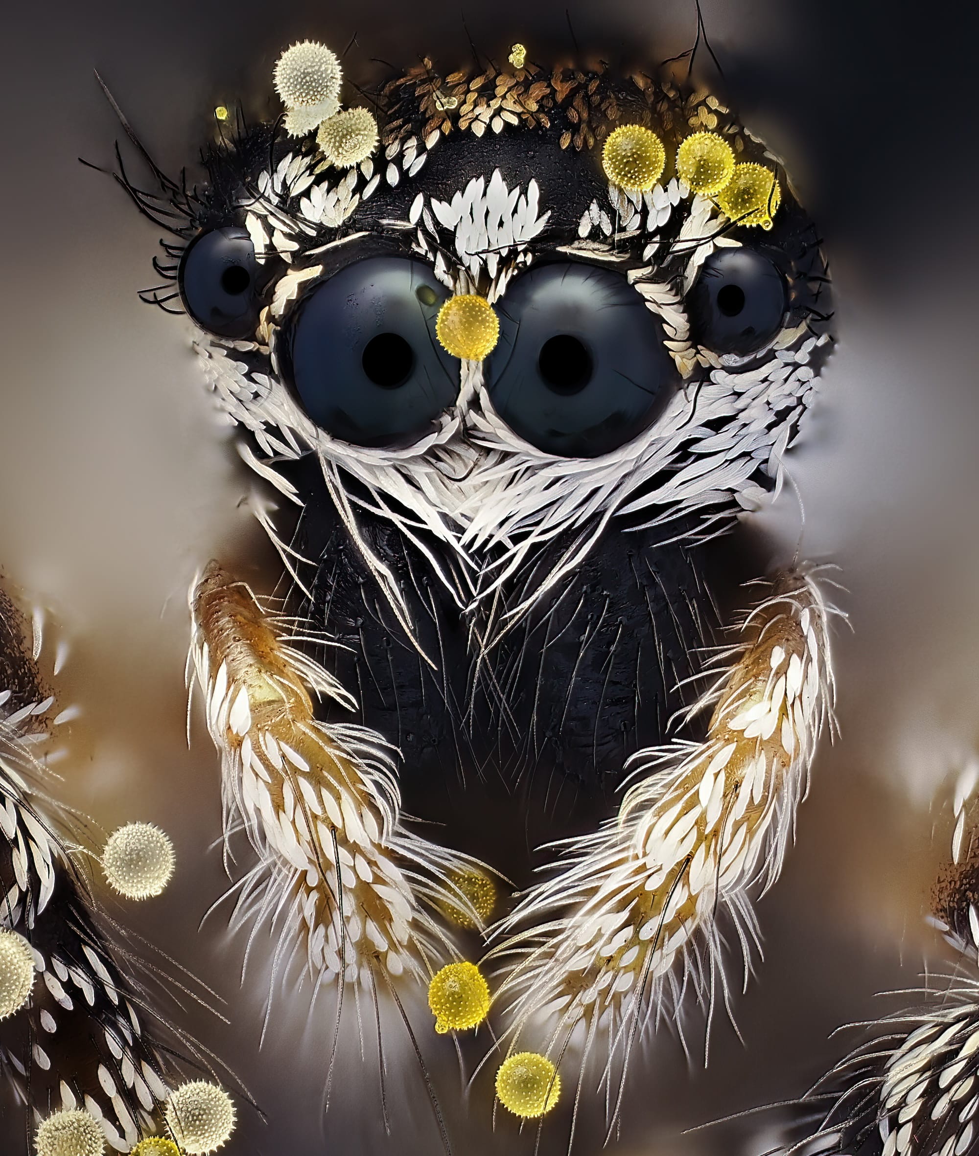
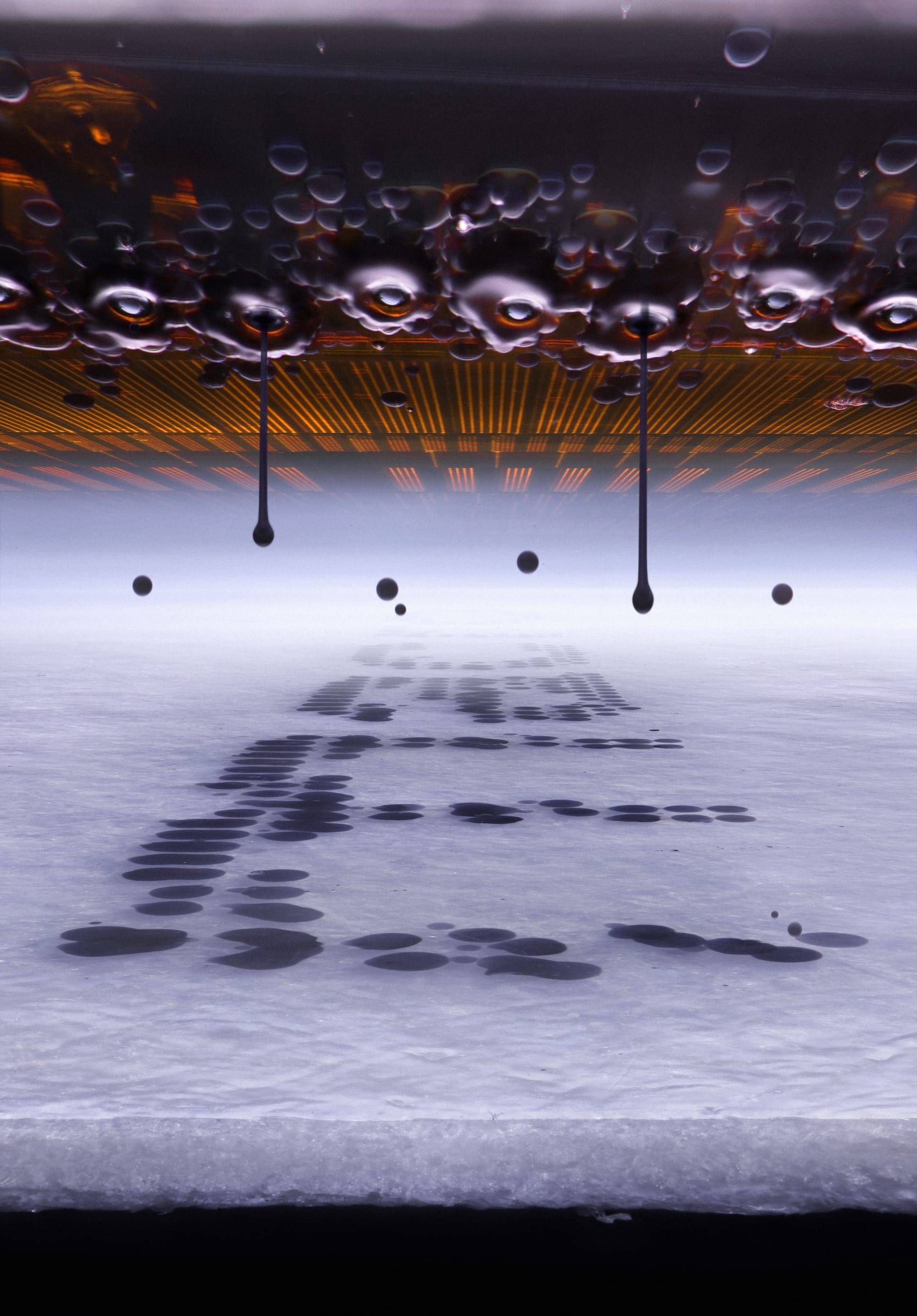
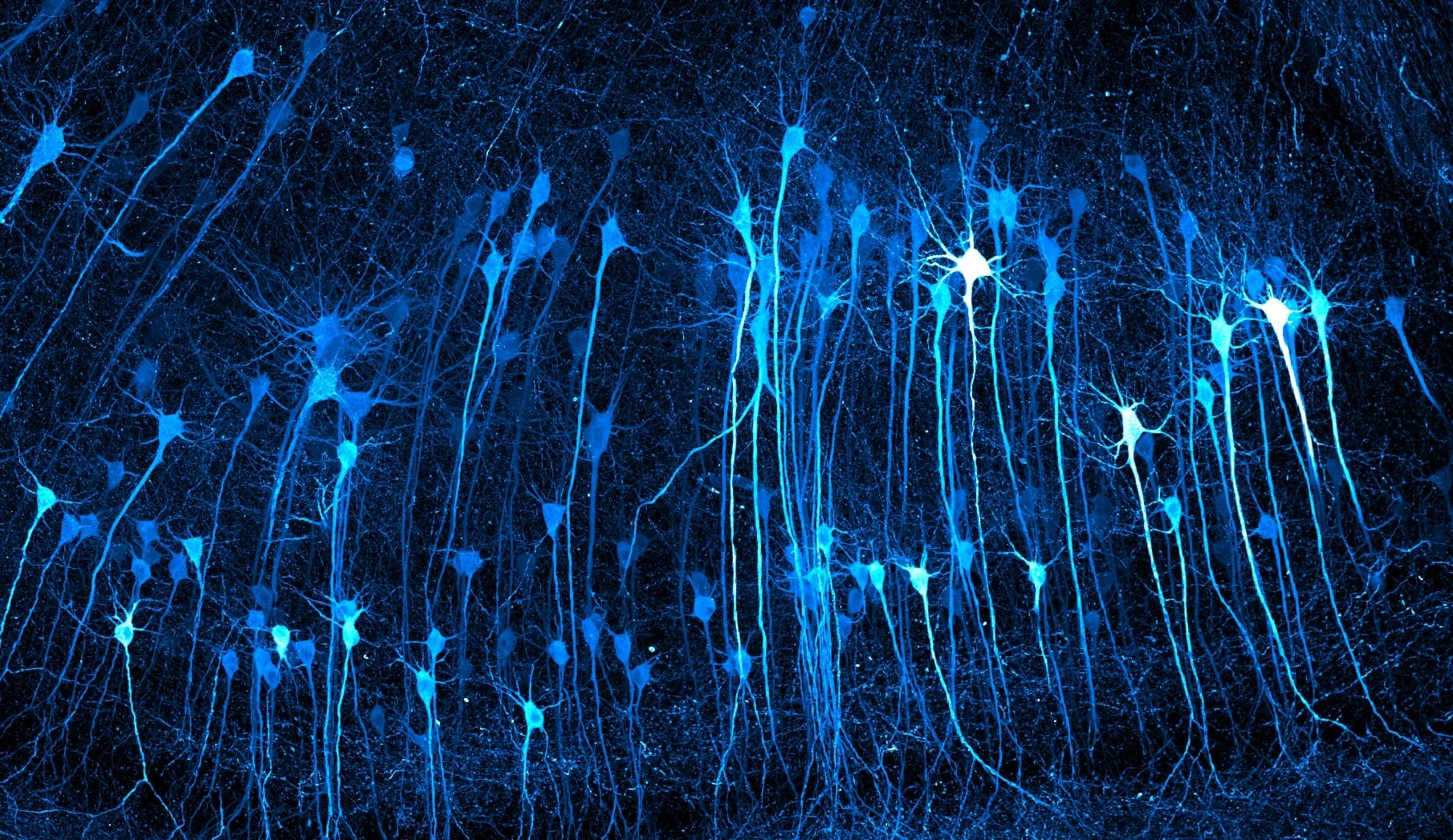
From fungi and insects to technology and crystals, the winning entries in the 2026 Nikon Small World Photomicrography Competition reveal astonishing details typically invisible to the naked eye. This year’s top prize went to Ole Bielfeldt, a student and experimental photographer based in Cologne, Germany, who used a hand-built rig to capture an incredible up-close view of droplets inside of an inkjet printer. Runners-up included John-Oliver Dum’s portrait of a jumping spider with pollen on its body and Kaelin Wulf and Rachel Flannery’s capture of mouse neurons.
Winner images were organized into three categories: Top 20, Honorable Mentions, and Images of Distinction. Explore some of our favorites here, and head to the contest’s website to see all of this year’s best photos. You might also enjoy the Nikon Small World in Motion video competition.
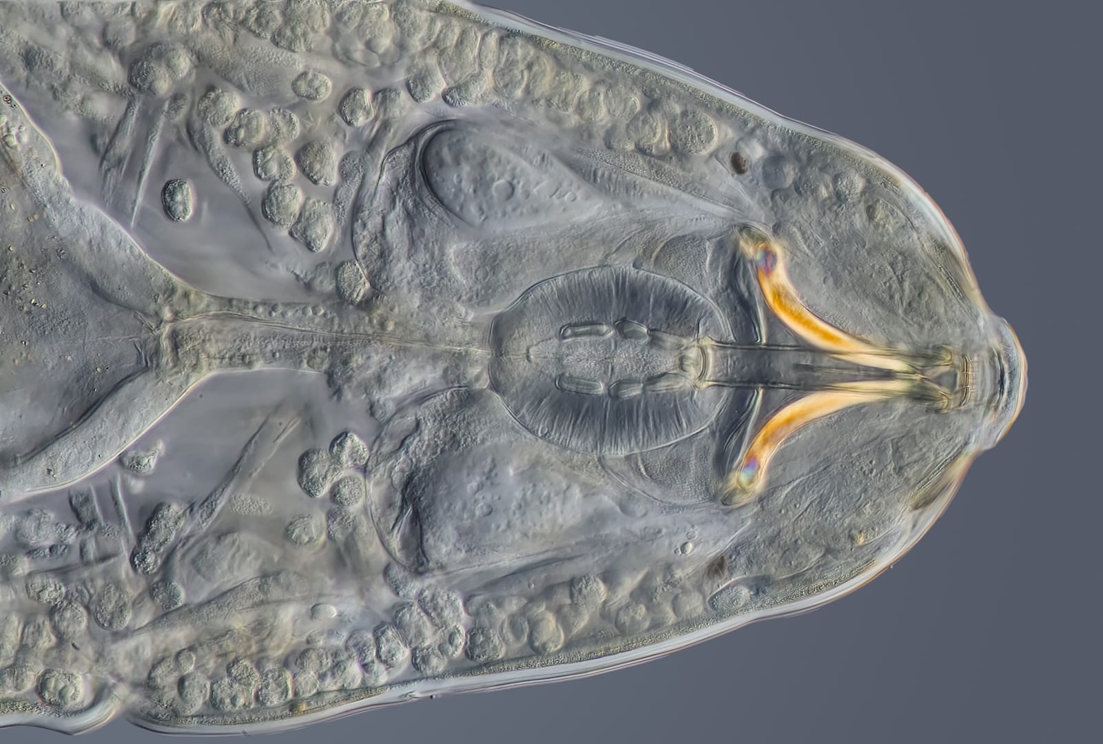
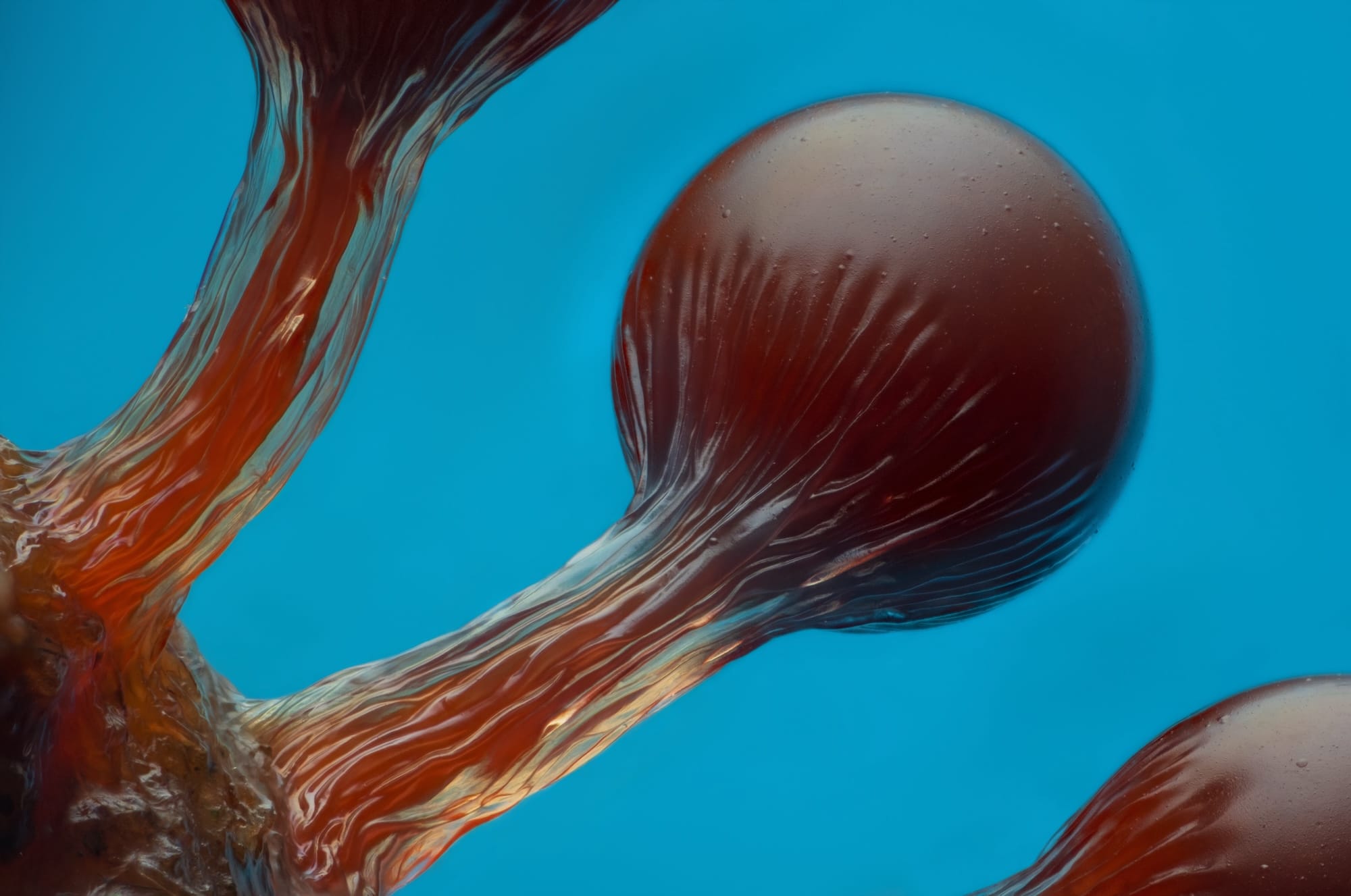
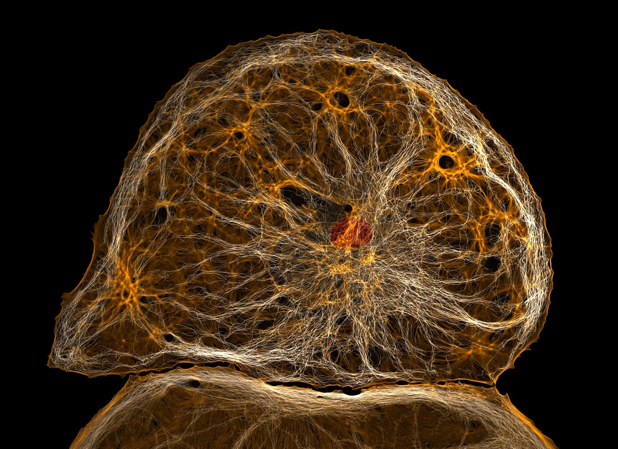
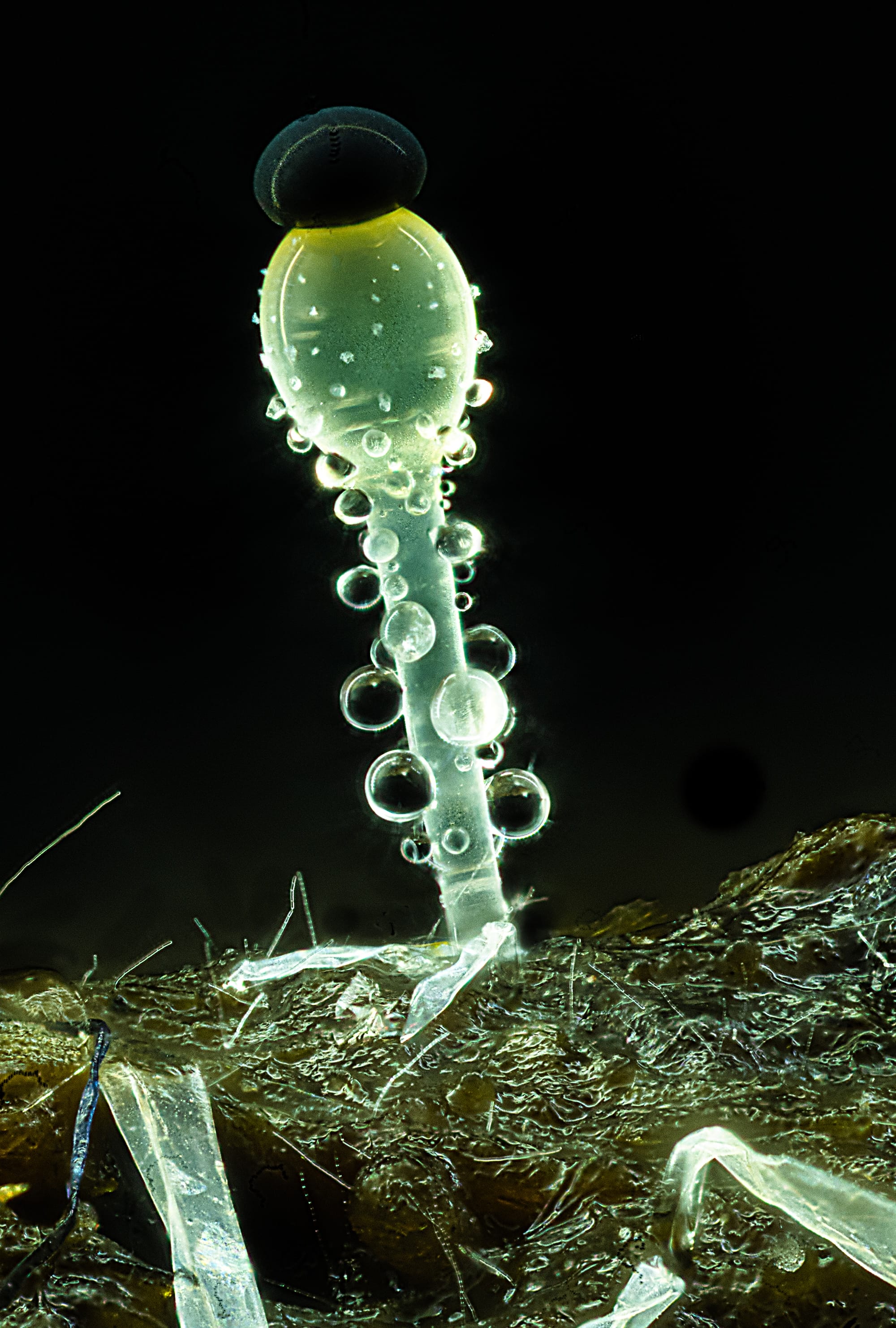
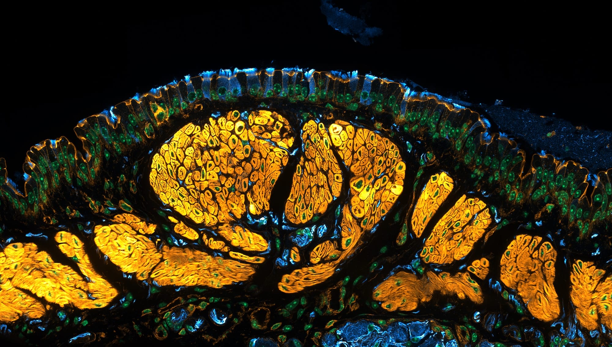
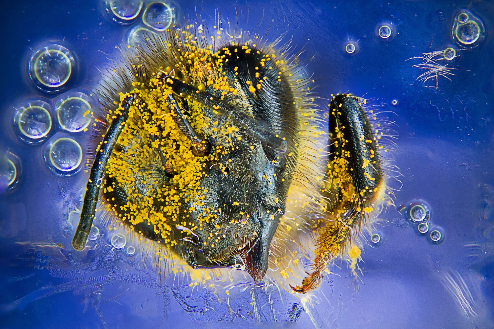
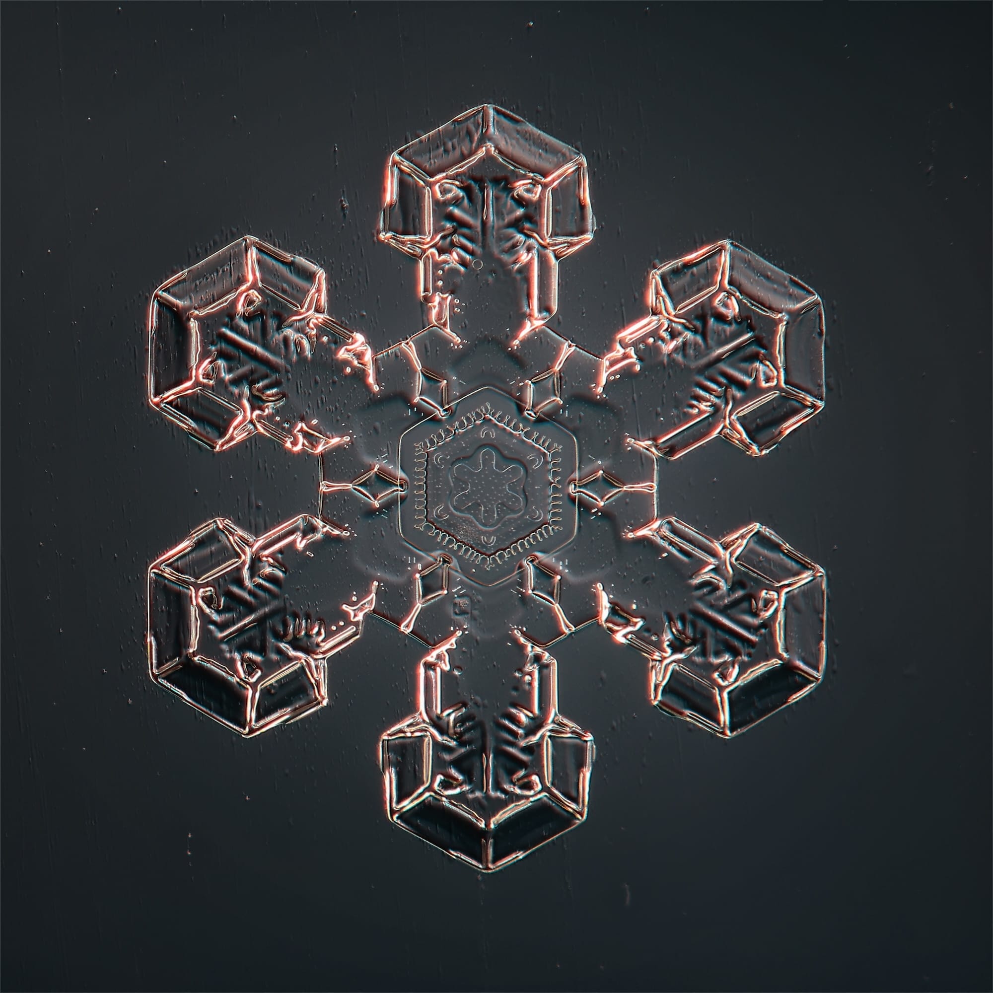
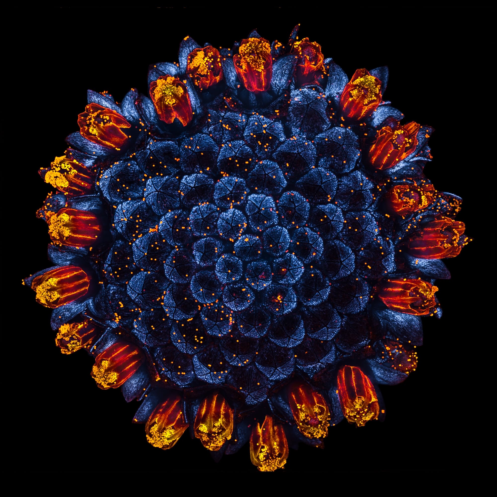
1st Place: Ole Bielfeldt (Cologne, Northrhine-Westfalia, Germany). Microscopic view of an inkjet printer depositing ink onto paper3rd Place: Kaelin Wulf and Ms. Rachel Flannery (Allen Institute, Department of Neural Dynamics, Seattle, Washington, U.S.). Neurons in an expanded mouse brainImage of Distinction: Charles Krebs (Issaquah, Washington, U.S.). Tardigrade (“Water Bear”) clearly showing the piercing stylets and pharnyx used to eat6th Place: Eric Epstein (Brooklyn, New York, U.S.). Hemitrichia slime mold spore structures (fruiting bodies)9th Place: Dr. Francisco Lázaro-Diéguez (Albert Einstein College of Medicine, Bronx, New York, U.S.) Dedifferentiated liver endothelial cell14th Place: Mike Crutchley (Pembrokeshire, Southwest Wales, U.K.). Pilobolus fungi on sheep dung15th Place: Dr. Julien Resseguier (University of Oslo, Department of Biosciences, Oslo, Viken, Norway). Mouse trachea showing tiny hair-like cells that move mucus and nearby nerves (blue) associated with the muscles (yellow)Image of Distinction: Giuseppe Franchina (Messina, Italy). Western honey bee (Apis mellifera) with pollenImage of Distinction: Sergey Kichigin (Vologda, Russia). A snowflake under a microscopeHonorable Mention: Jan Martinek (Czech Academy of Sciences, Institute of Experimental Botany, Prague, Czech Republic). A common daisy flower head (Bellis perennis) glowing with its natural autofluorescence